
 The 1,750 square foot MRI research facility is located on the first floor of the MRI Building. It is operated by dedicated MR registered technologists during normal business hours. The technologists have experience in all aspects of MR imaging research as well as with a wide variety of animal imaging. The facility is equipped with all necessary supplies, including resuscitation equipment.
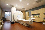
Siemens Magnetom Cima.X 3T MRI
Two Siemens Magnetom Cima.X 3T magnetic resonance whole body scanners are located on the first floor of the MRI Building. It possesses the strongest ever gradient system for a clinically released whole body MR, making smaller structures in the body visible, and capturing images faster than previous MR scanners. The Magnetom Cima.X has new features designed to enhance scientific research and overcome key imaging challenges in the visualization of cancer and other diseases. The 3T Cima.X is currently operating software version XA61A-SP01.
At the heart of the Magnetom Cima.X are its industry-best Gemini Gradients. These gradients have an amplitude of 200 mT/m and a slew rate of 200 T/m/s. These represent the strongest gradient system of any clinically available whole body MR scanner. The whole-body gradient coils bring significant improvement to diffusion weighted imaging, enabling the visualization of underlying structures that were previously unseeable.
The Cima.X delivers excellent homogeneity, a large 55 x 55 x 50 cm FOV, with a 2050 mm scan range and a dedicated network of sensors in the magnet allow constant adjustment of the transmit and receive parameters for maximum SNR.
The Cima.X features the next generation of Deep Resolve, which is an AI powered reconstruction technology that takes advantage of convolutional neural networks to accelerate MRI scans. Simultaneous Multi-Slice helps cut exam times, reduces DWI scan times throughout the body and significantly improves temporal resolution for BOLD fMRI. Compressed Sensing drastically reduces acquisition times and offers new capabilities for 3D imaging.
Magnetom Cima.X will allow physiologging of time stamped physiology signals for fMRI data correction. This makes a difference in precision by simplifying the correlation of physiology and fMRI data.
With a bore size of 60cm, and magnet length of 172cm the Cima.X provides ample comfort and FOV coverage for a broad range of exams.
The Cima.X comes with a wide variety of coils to meet the needs of both research and clinical applications, including BioMatrix head/Neck 20 channel, 32 Channel Head, 64 Channel Head/Neck, BioMatrix Spine 72 with respiratory sensor, Body 18, Ultraflex Small and Large 18, Tx/Rx Knee 18 and Body 30.
Software packages such as Neuro fMRI/DTI, SWI, Neuro Perfusion, PCASL, ASL 3D, ASL 2D, LiverLab, QISS, MyoMaps, SVS, and Flywheel provide a robust selection of options for research scanning. Also included is an fMRI Trigger Converter for external stimulation devices in fMRI experiments. The signal can be converted to an electrical signal (TTL/BNC and RS 232 interface for PC; modes: toggle or impulse). The Advanced cardiac package contains special sequences and protocols for advanced cardiac imaging including 3D and 4D BEAT functionalities. It supports advanced techniques for ventricular function imaging, dynamic imaging, tissue characterization, coronary imaging and more.
In addition to the software packages above, our second Cima X scanner installed in 2026 includes Multinuclear Spectroscopy capabilities. The hardware and software package required to prepare the MR system for spectroscopy and nuclear imaging with the nuclei 3He, 7Li, 13C, 17O, 19F, 31P, 23Na, and 129Xe is included. A Tim Coil interface MNO for connecting multi-nucleus coil is included. Coils with preamplifiers and transmitter duplexers as well as optimized pulse sequences for the individual nuclei are not included. In addition, a CSI spectroscopy package with 7Li, 13C, 17O, 19F, and 31P nuclei is available.
All image data are supplied to investigators in compliance with IRB and HIPAA regulations. Data can be downloaded to dedicated research image workstations for investigator analysis and/or to be formatted for transmission to offsite reading centers.
All image data is supplied to investigators in compliance with IRB and HIPAA regulations. Data can be downloaded to dedicated research image workstations for investigator analysis and/ or to be formatted for transmission to offsite reading centers.
Siemens MAGNETOM Skyra 3T MRI Scanner with TIM Technology
- 3T Siemens Skyra operating at E11E platform
- Multinuclear spectroscopy
- Gradient field strength of 45 mT/m, SR 200 T/m/s
- 70 cm open bore design, weight limit 500 lbs
- Capable of Advanced DTI, BOLD, Spectroscopy, ASL, Map-It (cartilage) and Advanced Cardiac Imaging
- Equipped with stimulation equipment for fMRI studies
- Various coils including 32 channel head coil, 20 channel head/neck, 18 channel body, 32 channel spine, 15 channel knee and surface loop coils.
- Contrast Power Injector